claire atkinson
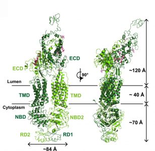
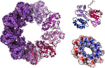
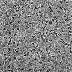
Structure of a lipid flippase from the retina from the Molday and Van Petegem Labs
Molday and Van Petegem Labs
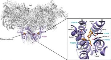
Structural basis of insecticide binding to the ryanodine receptor from the Van Petegem Lab
In a new paper from the Van Petegem lab, researchers show how the widely used diamide class of insecticides bind to an ion channel called the ryanodine receptor, and how the insecticide triggers opening of this ion channel. The study shows how several insects have developed resistance by evolving mutations directly within the binding site. […]
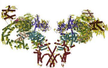
Structure EspA filament of enteropathogenic E.Coli from the Strynadka Lab
In a new paper from the Strynadka lab, researchers show the structure of the EspA filament from enteropathogenic E.Coli. Enteropathogenic E.Coli used the a multi-protein assembly called the Type III Secretion System to deliver proteins to host cells. The EspA filament is an extension to the Type III secretion system required for bacterial colonization of […]
Structure of EscV, a key protein in bacterial secretion systems from the Strynadka Lab
A new paper from the Strynadka lab uses cryo-EM to solve the structure of the bacterial protein EscV. EscV is a key protein in controlling the export of substrates through a macromolecular ‘syringe’ known as the Type III secretion system, which bacteria use to transfer proteins, including toxins, directly into host cells.
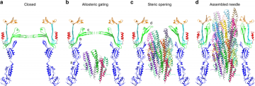
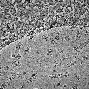
Near Atomic Cryo-EM Structure of the Type III Secretion System from the Strynadka Lab
Congratulations to the Strynadka Lab on their paper published in Nature Communications! Researchers used cryo-EM to determine a high resolution structure of the Type III Secretion System, and processed the data to obtain near atomic resolution structures of several components which make up this assembly.
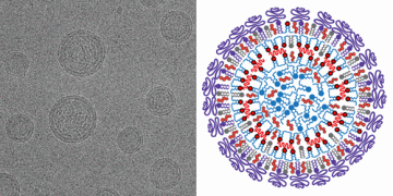
Lipid Nanoparticles from the Cullis Lab
Congratulations to the Cullis lab on their paper published in ACS Nano. Kulkarni et al used the HRMEM facility to image lipid nanoparticles.
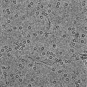
Apoferritin
Image of apoferritin captured on the Krios microscope at HRMEM.
Beta Galactosidase
Image of beta galactosidase captured on the Krios microscope at HRMEM.
Keyhole Limpet Haemocyanin
Image of keyhole limpet haemocyanin take on the Krios microscope at HRMEM.